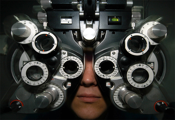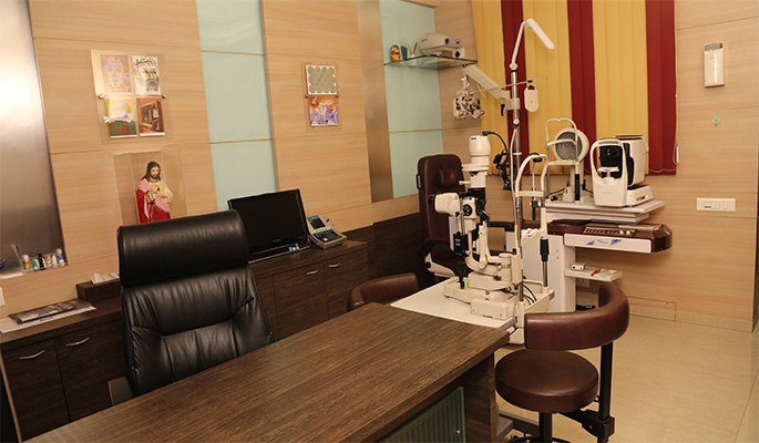
A complete routine eye examination, including a computerized refraction with a digital phoropter, eye pressure recording and a slit lamp examination for anterior and posterior segment evaluation is carried out. During this examination, the eye is examined for common eye problems like a refractive error and the possibility of a cataract, glaucoma and retinal diseases, among other conditions is ruled out.
CEDS Eye Hospital is well equipped with all the necessary instruments to diagnose and institute the first line treatment of all common eye diseases. It offers Comprehensive Ophthalmology services that include a full range of medical and surgical eye care. When a medical condition so indicates, the consultant will refer the patient to the care of the appropriate Ophthalmology sub-specialist.

The patient is first examined by highly qualified Doctors of Optometry for refractive errors and related conditions using a Huwitz Automated Digital Optometry System in the Optometry Chamber which ensures a smooth and automatic assessment of Visual Function.
The optometrist then prescribesthe relevant optical correction and will refer the patient to our in- house Opticians for dispensation or the patient may seek his/ her own Optician.
Anterior Segment evaluation is then carried out with Digital Slit Lamp Bio Microscope followed by recording of intra-ocular pressure with the help of Applanation Tonometry or Non-Contact Pneumatic Tonometry
Dilation of the pupil, if necessary, is done and then retinal evaluation is carried out using an indirect ophthalmoscope or a +90 Diopter lens.
After this baseline examination, additional diagnostic procedures like Corneal Topography, Wavefront Aberrometry, Optical Coherence Tomographymay be ordered at the discretion of the examining doctor, based on the patient’s specific requirements.
At the end of this examination, appropriate medication and / or optical aids like spectacle glasses or contact lenses are suggested to the patient. A treatment plan and guidelines for further management and review will be presented to the patient for consideration, where indicated.

AUTOMATED REFRACTION UNITS: A Hewitz Automated Optometry System is installed in the Optometry Chamber which ensures a smooth and automatic assessment of Visual Function during Preliminary Examination by the Optometrist. The Optometry Unit also possesses a Humphrey Automated Refraction Unit from Carl Zeiss, a TOPCON VISION CHART Projector and a TOPCON Automated Lens Meter for the accurate verification of a patient’s existing spectacle or contact lens prescription. A Manual Phoropter System from Topcon, Japan is also present as a backup measure.
VECTOR VISION CONTRAST SENSITIVITY TESTING SYSTEM: The Center is equipped with the Vector Vision Contrast Sensitivity Testing system, one of the very few installations in the country, confirming to modern standards of Visual Assessment. This modality of vision testing is fast gaining acceptance as the new gold standard in the qualitative assessment of vision in patients with Glaucoma, Cataracts, Diabetic Eye Disease and following the implantation of New Generation Multifocal Lens Implants.
EYE EXAMINATION UNIT: A single point stop for the patient to be examined for most Baseline and Routine Eye Examination
TOPCON SLIT LAMP BIOMICROSCOPE WITH IMAGING SYSTEM: The Center is equipped with a high end Digital Slit Lamp Biomicroscope from TOPCON, Japan, which can help the doctor to accurately observe and diagnose diseases afflicting the eye. In addition, the Slit Lamp is equipped with an Image Acquisition System which can allow relatives of the patient to simultaneously see and understand the eye as the Consultant examines the patient. Images from the patients eyes can then be stored for future reference (a valuable tool in assessing the progression of any disease), retrieved at will, printed as a hard copy or emailed to the patient for purposes of records. These images can also be used for scientific research and publications in the future if required (with the explicit written permission of the patient).